Introduction
Obstructive sleep apnoea syndrome is the result of upper airway obstruction from airway narrowing and closure during sleep. It is associated with increased cardiovascular morbidity and mortality. The manifestations of obstructive sleep apnoea, such as excessive daytime sleepiness, daytime fatigue and poor sleep quality due to sleep fragmentation, are also well established. Currently, nasal continuous positive airway pressure (CPAP) is the first line of treatment. The effectiveness of nasal CPAP in improving the symptoms of obstructive sleep apnoea is irrefutable. A detailed nasal exam with endoscopy is critical in evaluating a patient for nasal CPAP (Unfortunately this is rarely done by most physicians assessing and managing patients with sleep apnoea). Nevertheless, patient compliance and tolerance represents a clear problem. Furthermore, even in compliant patients who are using CPAP on a "regular basis," the actual usage is only approximately 50% of the ideal. Due to the limitations of CPAP, surgical treatment of OSA should be considered as a viable treatment option. Indeed, most of the patients Dr Broadhurst operates on are unable to use CPAP for various reasons and so clearly need other options to manage their sleep apnoea.
This review will present the current state of art in sleep apnoea surgery, beginning with evaluation of the patient as surgical candidate to formulation of a surgical plan through procedural selection based on published surgical outcomes.
Pre-Operative Assessment
Numerous surgical procedures are currently available for the treatment of obstructive sleep apnoea. However, the following issues present a formidable challenge to the sleep apnoea surgeon:
- the complex interplay of the soft and hard tissues that contribute to upper airway obstruction
- the crucial role of this anatomic region to speech and swallowing
- the subsequent oedematous response after surgical intervention.
Moreover, it is well accepted that successful surgical outcomes depend on proper patient selection as well as the choice of surgical procedure(s). Therefore, a logical and systematic approach to clinical evaluation, treatment planning, surgical execution and perioperative management is necessary to maximize safety and improve outcomes.
Clinical evaluation must include the overall body habitus (height, weight), since it has been shown that surgical outcomes can be influenced by these factors. A detailed examination should focus on the head and neck region in order to identify the potential sites of upper airway obstruction, including the nose, soft palate, lateral pharyngeal walls and tongue base. This is best performed by an ENT (ear, nose throat) surgeon trained in managing sleep apnoea. The presence of items listed below are common findings in patients with obstructive sleep apnoea:
- nasal septal deviation
- turbinate hypertrophy
- nasal valve collapse
- elongation of the soft palate/uvula
- tonsillar hypertrophy
- enlargement of the tongue
- narrowed and/or deficient maxilla and mandible
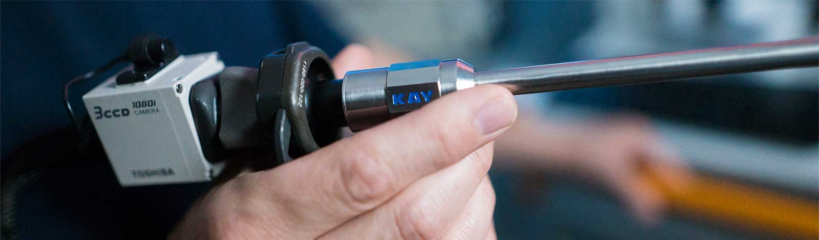
An upper airway examination by a fibre-optic or rigid telescope is highly recommended in patients with obstructive sleep apnoea. This evaluation enables Dr Broadhurst to directly visualise the entire upper airway from the nose to the larynx. The dimension of the nasal, velopharyngeal and hypopharyngeal airway can be fully assessed. Furthermore, the prominence of the tongue base and the lateral pharyngeal wall, as well as their collapsibility, can be evaluated with the Muller's maneuver. Dr Broadhurst uses the Friedman classification of tongue position to assess the oropharyngeal airway. With this comprehensive examination, the upper airway can be completely assessed for anatomic abnormalities that may be contributing to obstructive sleep apnoea. Directing surgery to specific multi-level obstruction is the key to success.
Surgical Procedures
The key for the surgical planning is selecting the appropriate surgical options for the levels of obstruction in the nose, mouth and throat. The most common combination for Dr Broadhurst is a septoplasty with reduction of the inferior turbinates together with an anterior palatoplasty (+/- tonsillectomy) and radiofrequency reduction of the tongue base. Below are descriptions of the various procedures described in the sleep apnoea surgery literature, not all of which Dr Broadhurst uses or feels are good choices.
Nasal Surgery
The relationship between nasal obstruction and sleep-disordered breathing has been demonstrated by numerous investigations. Both daytime nasal obstruction and nocturnal nasal congestion have been shown as risk factors for sleep-disordered breathing. Therefore, the treatment of nasal obstruction plays an important role in sleep apnoea surgery. However, it must be emphasized that although obstructive sleep apnoea can be improved in some patients, only slight improvement has been shown with nasal surgery alone. Three anatomic areas of the nose that may contribute to obstruction are the septum, the inferior turbinates (IT) and the nasal valve region. The most common nasal surgical procedure consists of a septoplasty and turbinate reduction. The major effects of nasal surgery are subjective improvement of nasal patency and reduction of the nasal CPAP requirement. Many patients also notice an improvement in snoring with this surgery alone and Dr Broadhurst uses the septoplasty combined with radiofrequency reduction of the inferior turbinates as his treatment of choice. This is the current teaching of world leaders in this field and represents the most up to date technique.
Other techniques exist for reduction of the IT including submucosal shaving, cautery to the surface and physical removal of the turbinates. The radiofrequency procedure is felt to be highly efficient and has the least morbidity compared with other techniques and so is Dr Broadhurst’s preferred choice for IT surgery.
Uvulopalatopharyngoplasty
Uvulopalatopharyngoplasty (UPPP) has been the most common sleep apnoea surgical procedure performed during the past 25 years. Traditional UPPP procedure consists of the removal of redundant soft palate and pharyngeal tissues as well as uvula, to widen the oropharyngeal inlet. The tonsils are also removed if present. Although UPPP can improve oropharyngeal obstruction, hypopharyngeal obstruction is minimally improved by the procedure, thereby reflecting a success rate as low as 40%. Furthermore, potential complications including velopharyngeal insufficiency (water leaking up into the nose), stenosis and dysphagia are major concerns. Patients also complain of a very dry and sore throat as the function of the uvula are now gone.
Consequently, several modifications of the traditional procedure have been developed to improve outcomes and reduce complications. Of these, Dr Broadhurst feels the anterior palatoplasty is the most effective. This is a technique taught by many world leading sleep apnoea surgeons and is the current best-accepted technique. It involves removal of a rectangle of soft palate up hear its junction with the hard palate and retains the uvula and free edge of the soft palate. This preserves the normal palate function and patients do not report velopharyngeal insufficiency (water leaking up into the nose), stenosis, dysphagia or a very dry and sore throat. Currently in Australia, there are only few surgeons using these new and advanced techniques. As with many areas in surgery, most surgeons tend to be quite slow to adopt new techniques even if they have a well-proven track record of improving outcomes.
Other Palate Procedures
Uvulopalatal Flap
The uvulopalatal flap procedure is a modification of UPPP, which results in the widening of the oropharyngeal airway by suspension of the uvula superiorly toward the hard-soft palate junction after a limited resection of the uvula, lateral pharyngeal wall and the mucosa. As such, this surgical technique results in the widening of the oropharyngeal airway. A prospective study of 80 patients with variables including age, sex, body mass index, soft palatal length, respiratory disturbance index (RDI), lowest oxygen desaturation (LSAT) and subjective snoring scale demonstrated that there was no statistical significant difference between the uvulopalatal flap group and the UPPP group. Using a visual analog scale to assess pain, there was significantly less pain in the uvulopalatal flap group vs. the UPPP group. This remains an out-of-favour procedure as with the laser assisted uvulopalatoplasty or LAUP (see below) and UPPP.
Pharyngoplasty
The pharyngoplasty procedure is another modification of the UPPP. This procedure usually involves the removal of tonsillar tissues, but the uvula and the soft palate is preserved. The pharyngeal inlet is widened and the airway collapsibility is reduced purely via suturing of the tonsillar wounds. This procedure represents the most conservative pharyngeal surgery and has minimal side effects as compared to other procedures. This is not commonly done.
Laser-Assisted Uvulopalatoplasty
Laser-Assisted Uvulopalatoplasty (LAUP) was introduced by Kamimi as an office-based surgical procedure for the treatment of snoring. The procedure involves removal of the uvula and a portion of the soft palate by carbon dioxide laser incisions and vaporization. Most of the uvula is amputated, and the soft palate (1-2 cm lateral to the uvula) is incised and vaporized. Additionally, mucosal or tonsillar pillar tissue is vaporized as needed.
Although many studies have evaluated the efficacy of LAUP in the treatment of OSA, most of the studies are flawed by methodological discrepancies or statistical inadequacies such as ill-defined criteria for response and lack of adequate follow-up. In addition, LAUP is associated with increased risk of dysphagia as well as the risk of discontinuation of treatment due to pain. Moreover, recent AASM evidence-based guidelines do not support the use of LAUP in the treatment of obstructive sleep apnoea and so should be strongly avoided.
Temperature-Controlled Radiofrequency Tongue Base Reduction
Low-wave radiofrequency energy achieves therapeutic ablation of tissue in a minimally invasive fashion. Radiofrequency can be safely applied to the upper airway tissue to improve OSA by volumetric tissue reduction and tissue stiffening.
Radiofrequency energy delivery to the upper airway tissue occurs through a needle electrode. The energy current causes ionic agitation of the tissue around the electrode, resulting in frictional heating of the tissue. Therefore, the electrode itself does not get hot; heat actually emanates from the tissue. Tissue injury occurs when the temperature reaches beyond 47°C, which is when cell proteins undergo denaturation. The size of the lesion (area of tissue injury) created in the tongue base is dependent on the current intensity and the duration of energy delivery. Typically, the lesion is in the shape of an ellipse.
Since the radiofrequency energy disbursement is proportional to 1/radius4, heat dissipation is limited and excessive tissue injury is minimized. Furthermore, when the temperature reaches 90°C, char formation on the electrode leads to an increase in impedance and results in disruption of current flow, thus serving as a second layer of protection. These factors allow radiofrequency ablation to create a predictable tissue injury pattern, thereby minimizing potential complications.
The first prospective study of radiofrequency tongue reduction was conducted in 18 patients. After a mean total energy of 8490 joules was delivered per patient over a mean of 5.5 treatments, the mean RDI improved from 39.6 to 17.8 with an improved LSAT from 81.9% to 88.3%. The tongue volume was reduced by a mean of 17% (based on MRI findings). There were no changes in speech or swallowing. Complications included a superficial tongue ulceration that resolved spontaneously, persistent pain on swallowing that resolved after several weeks, and a tongue abscess that required drainage. Sixteen of the 18 patients were followed on a long-term basis (mean of 28 ± 4 months). Findings demonstrated a mean weight increase of 3.1 ± 7.9 kg. In addition, there was a worsening of RDI from 17.8 to 28.7. There was also a worsening trend in the LSAT from 88.1% to 85.8%. However, there was no significant deterioration of the quality of life measurements by SF-36 or daytime sleepiness by Epworth Sleepiness Scale (ESS).
Multiple other reports have shown radiofrequency tongue reduction to be efficacious in improving obstructive sleep apnoea. However, based on all reports, improvement with radiofrequency tongue reduction is insufficient as a single treatment modality. Therefore, Dr Broadhurst uses it as an adjunctive treatment in combination with other surgical approaches to maximize the patient outcome.
Advanced Procedures for Severe Abnormalities
The following procedures are substantially invasive and not performed routinely by Dr Broadhurst. They are performed by very few surgeons in the country and if it is felt necessary, then Dr Broadhurst will discuss this with you and arrange a review by a suitable surgeon.
Genioglossus Advancement
The mandible and the tongue are major determinants of the airway dimension. Anterior positioning of these structures has been shown to improve obstructive sleep apnoea. The genioglossus advancement procedure is limited to moving forward the geniotubercle with the genioglossus insertion without moving the mandible. This advancement places tension on the tongue musculature, thereby limiting the posterior displacement during sleep. The genioglossus advancement procedure consists of a rectangular osteotomy on the symphysis of the mandible intraorally. The rectangle is advanced forward by the thickness of the mandible and partially rotated to prevent retraction back into the floor of the mouth. Incorporation of the geniotubercle during the procedure has been shown to be quite successful with this technique. In general, genioglossus advancement is performed with other sleep apnoea surgical procedures such as UPPP and hyoid advancement in order to maximize improvement. The results from these procedures have been variable, ranging from 23% to 77%. These variable results underline the difficulty in accurately predicting the success rate. Clearly, anatomic factors, body habitus and obstructive sleep apnoea severity are all factors that influence surgical success. In general, the potential risks associated with genioglossus advancement are quite limited, including infection, hematoma, injury to the genioglossus muscle and paresthesia of the lower teeth.
Hyoid Advancement
The hyoid bone holds an intimate relationship with the tongue base and pharyngeal musculature, thus portraying an integral aspect of the upper airway anatomy. The hyoid bone may be surgically repositioned anteriorly by attaching it to the thyroid cartilage in order to expand the airway. The procedure is usually performed in conjunction with genioglossus advancement so as to contribute to the improvement of obstructive sleep apnoea. However, some surgeons have elected to combine it with UPPP alone. An inherent problem with hyoid advancement is the requirement of an external incision on the neck, one aspect that may not be readily accepted by all patients. As with other sleep apnoea surgical procedures, the results of hyoid advancement are variable, ranging from 23% to 65%. In general, the associated surgical risks are low, and may include infection, seroma formation and dysphagia.
Maxillomandibular Advancement
Abnormality of the maxillofacial skeleton is a well-recognized risk factor of obstructive sleep apnoea. Maxillomandibular advancement was initially advocated based on the finding that maxillofacial skeletal abnormality (i.e., maxillary and/or mandibular deficiency) is frequently found in patients with obstructive sleep apnoea, and that maxillomandibular deficiency results in diminished airway dimension, which leads to nocturnal obstruction. Maxillomandibular advancement achieves enlargement of the entire upper airway including the nasal, pharyngeal and hypopharyngeal airway expansions of the skeletal framework that encircle the airway. Comparison of pre- and post-operative airway appearance based on fibre-optic nasopharyngoscopy and lateral cephalometric radiograph have demonstrated that in addition to airway expansion by the forward movement of the maxillomandibular complex, the tension and collapsibility of the suprahyoid and velopharyngeal musculature may also be reduced, thus leading to the reduction of lateral pharyngeal wall collapse.
The maxillomandibular advancement procedure consists of mobilizing the maxilla and mandible to achieve anterior displacement of the maxillomandibular complex after intraoral osteotomy of the maxilla and mandible. The maxilla and mandible are stabilized with titanium plates in the advanced position. In order to maximize the airway expansion, an advancement of 10-12 mm is usually recommended. However, it is important to achieve maximal advancement while maintaining a stable dental occlusion as well as balanced aesthetic appearance. Interestingly, although many patients may be left with "prominent jaws," very few of them are dissatisfied with their appearance.
Maxillomandibular advancement is the most effective sleep apnoea surgical procedure currently available. The success rate is usually between 75% and 100% with a long-term success rate approaching 90%. In addition, patient perception of the surgical outcome has been very favorable. Although maxillomandibular advancement is considered a fairly invasive procedure, the associated surgical risks are low, including bleeding, infection, malocclusion and permanent numbness.
Maxillomandibular Expansion
A constricted maxilla with a high and narrowed hard palate contributes to increased nasal resistance and is a common finding in patients with obstructive sleep apnoea. Investigators have demonstrated that expansion of the maxilla can improve obstructive sleep apnoea in children and adolescents as well as adults. Furthermore, since patients with maxillary constriction often have a corresponding mandibular constriction, expansion of the maxillary and mandibular complex has also been shown to be beneficial in reducing the severity of obstructive sleep apnoea. The procedure consists of limited osteotomies to allow widening of the maxilla and mandible with distractors. The advantage of maxillomandibular expansion is that it is considerably less invasive than maxillomandibular advancement. However, treatment time is lengthened and patients need to keep the distractors in place for several months after the operation to ensure that the expansion is stable. Therefore, patient acceptance for this treatment option may be affected. In addition, orthodontic therapy is also mandatory and may possibly influence patient acceptance of this treatment modality.
Tracheotomy
The use of tracheostomy to bypass upper airway obstruction in "Pickwickian" patients was the first reported treatment for obstructive sleep apnoea. Despite being the most effective treatment for obstructive sleep apnoea, patient acceptance is low due to the associated morbidity and social implications. The current use of tracheotomy primarily serves as a temporary measure for airway protection in patients with severe sleep apnoea with either morbid obesity or significant craniofacial anomalies that pose a high risk for airway compromise in the perioperative period. However, permanent tracheotomy as a long-term treatment of obstructive sleep apnoea remains an option in morbidly obese patients with obesity hypoventilation syndrome or in patients with significant craniofacial anomaly who have failed all other forms of non-surgical and surgical treatments.
Surgical Planning
Clearly, prior to any sleep apnoea surgery, the diagnosis of obstructive sleep apnoea based on sleep study results is essential. Although some may debate whether a formal sleep study should be mandatory, the use of an ambulatory sleep study is an acceptable practice under current standards. The selection of surgical procedure(s) is based on numerous factors (Table 1). The patient's desire and preference as well as the health status can clearly influence outcomes and must be taken into consideration. Additionally, the goal of surgery may be different for different patients. Although the majority elect surgical treatment due to intolerance of non-surgical treatments (CPAP), some patients may consider surgery in order to improve their ability to tolerate non-surgical treatments, such as the reduction of therapeutic CPAP pressure or improvement of nasal symptoms due to CPAP use. Therefore, the surgical endpoint should be discussed prior to surgery, along with criteria for surgical success. Informed consent must be conducted, and patients should be educated regarding the rationale of surgery as well as associated risks and benefits.
In formulating a surgical plan, the most important task for the surgeon is to decide which procedure(s) should be utilised. Information gathered from the pre-operative assessment including clinical examination and fibre-optic nasopharyngoscopy can provide useful information regarding the upper airway anatomy and the site(s) of obstruction.
Clearly, the most logical surgical approach would be to minimise surgical intervention and avoid unnecessary surgery while achieving a successful result. Therefore, the majority of surgeons including Dr Broadhurst have favored a staged surgical protocol. The anterior palatoplasty with tongue base reduction and IT reduction with septoplasty is what Dr Broadhurst selects this as the first surgical line to improve obstructive sleep apnoea. 3 months after surgery a post-operative sleep study is obtained to evaluate the outcome. In patients with persistent obstructive sleep apnoea, further soft tissue surgery may be suggested within the staged protocol or more advanced and major surgery may be considered.
Patients with severe obstructive sleep apnoea, morbid obesity or significant hypopharyngeal obstruction such as severe mandibular deficiency, or patients who wish to have the best chance for a cure with a single operation can certainly be considered as candidates for maxillomandibular advancement as a primary surgical treatment option.
Post Operative Period
It is very common for 2-3 weeks to have significant throat pain with swallowing. This is worst at the 1-week mark and then slowly improves. Regular pain relief is critical to “making it through” this period. You can eat what ever you feel comfortable with but if you notice frank blood in the mouth this can be potentially serious. Firstly, suck on an ice cube for 15 minutes and if the bleeding does not cease, proceed straight to an emergency department of a hospital that can manage post-surgery bleeding like this. Fortunately, this is uncommon. You may need to call the ambulance if the bleeding is significant.
If you have had nasal surgery as part of your OSA procedure, then twice-daily nasal washes are essential to good healing of the nose. This continue for at least 2 weeks as significant crusts can build up causing some nasal blockage. For 4 days only, you need to use twice daily Drixine or another over the counter nasal decongestant to each side of the nose. This helps keep the nose open as it heals. It is common to experience nasal discharge of blood stained mucous. Gentle nose blowing is perfectly fine to help with this. Dr Broadhurst will review at 2-3 weeks following your surgery.
Conclusion
Successful surgical outcome depends on proper patient selection as well as the choice of surgical procedure(s). The adaptation of a logical and systematic approach to clinical evaluation, treatment planning and surgical execution is necessary in order to maximize safety and improve surgical results. Many new surgical techniques and evolving technology offer less invasive treatment modalities now with broader patient acceptance and improved results and Dr Broadhurst maintains an active interest in this the ensure patients receive the most up-to-date approaches to managing obstructive sleep apnoea.
Table 1: Factors influencing sleep apnoea surgery outcomes
|
|
Favorable |
Unfavorable |
|
Age |
Younger patients (< 60yr) |
Older patients (> 60yr) |
|
Body habitus |
Non-obese |
Obese |
|
OSA severity |
Mild to moderate (RDI < 30) |
Severe (RDI > 30) |
|
Site of obstruction |
Oropharyngeal (with tonsils) |
Hypopharyngeal |